Hands pulling apart chromosomes as they separate in a cell

CREATORS – Beata Mierzwa

In ALL, CREATORS by McKenzie Prillaman

Name: Beata Edyta Mierzwa

Which came first in your life, the science or the art?

Although professionally I am a scientist first, I always loved drawing and have been creating art for as long as I can remember. I was lucky to grow up with a little bit of both worlds—my mother is an artist, and my father an engineer. When I was finishing high school, my three big interests were science, art, and fashion. But I wasn’t sure which one to pursue as my main career, thinking that these paths were mutually exclusive. I went on to study molecular biology, and it wasn’t until I was almost finished with my PhD research that I rediscovered my passion for art.

“My three big interests were science, art, and fashion. But I wasn’t sure which one to pursue as my main career, thinking that these paths were mutually exclusive.”

Beata Mierzwa

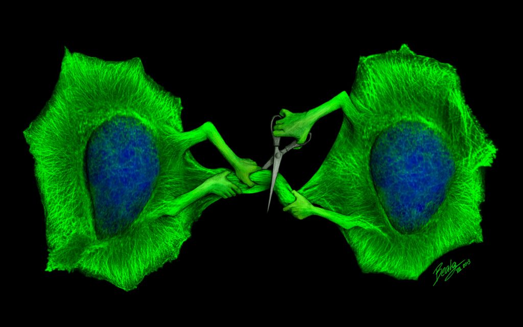
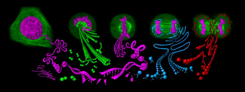
I created my first science illustration to depict the theme of my research on the final step in cell division—two cells literally cutting their connection with a pair of scissors. Although not a classical scientific figure, it was a fun and memorable way to present my research, and I was amazed to see that people would remember my findings years later. This motivated me to create more drawings for friends, colleagues, and other scientists, and I started making artwork for journal covers, conferences, and research groups. Spending time behind the microscope also allowed me to appreciate the beauty of cellular structures, which inspired me to create my own science fashion line using prints of visually stunning microscopy images. I would have never dreamed that one day I’d be able to combine my passions to communicate science in creative ways and spark fascination for biology.

Two bright green cells containing blue masses of DNA in their centers. The cells are connected in the middle, but the cells also have arms and are cutting their connection with scissors.
The Final Cut (2013) by Beata Mierzwa. In the final step of cell division, the bridge connecting the cells is cut in a process called cytokinetic abscission, and gives rise to two separate daughter cells. This hand-drawn illustration is overlaid with real microscopy images of a human cell, with the blue DNA carrying the genetic information and the green microtubules giving shape to the cell—a combination that quite literally fuses science and art!
Blue and orange cells with visible DNA. Brown arrows have pierced into the orange cells.
An Arrow Poison for CRISPR (2017) by Beata Mierzwa. This artwork illustrates a selection method for CRISPR/Cas9 gene editing. Cells that receive the desired mutation also become resistant to ouabain, a molecule traditionally used as an arrow poison in Africa, with its chemical structure highlighted on the feathers of the arrows. Cells without the mutation are struck by the arrows, leaving only cells that have successfully undergone genome editing. This drawing was featured on the cover of Nature Methods in June 2017.
A spiral of lime green and blue building blocks. Lego people are adding more Lego blocks to the spiral.
ESCRT Lego (2016) by Beata Mierzwa. This artwork illustrates the dynamics of a protein machinery for splitting membranes. This machinery, called ESCRT-III, forms spirals that constantly exchange their building blocks with the help of a protein called VPS4. This remodeling allows the spirals to change their shape and constrict membranes until they split in the final step of cell division to separate the emerging daughter cells.

Which sciences relate to your art practice?

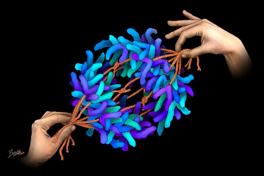
My illustrations focus on the tiny universe within our cells—depicting molecular processes that make all life possible, but in a way that can be appreciated both inside and outside the scientific community.

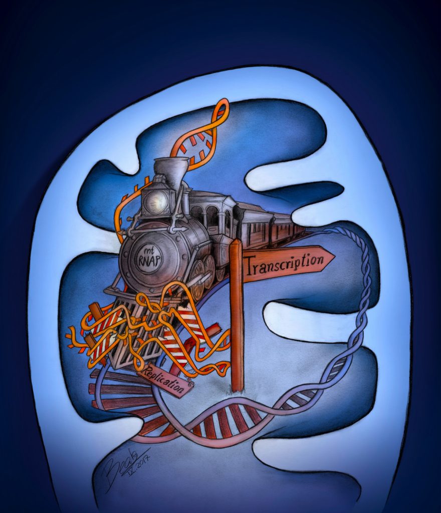
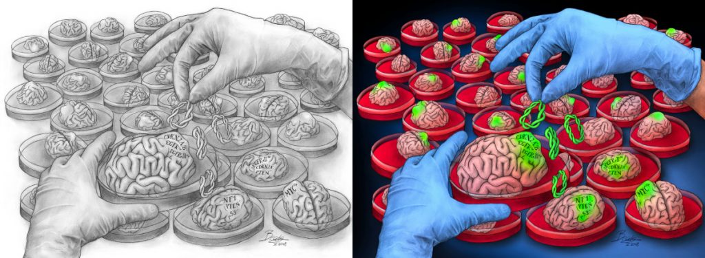
Many of my drawings reflect my fascination for cell division, a research field I’ve been passionate about ever since I started my master’s thesis. I also enjoy illustrating recent scientific discoveries from diverse fields—such as the intricate organization of cells in the brain, mechanisms of gene regulation, molecular processes inside mitochondria, or novel methods that revolutionize scientific research.

Within a tangle of pink DNA strands are two blue proteins in handcuffs and a turquoise protein unwinding a DNA strand and holding scissors to it.
Handcuffs for CRISPR (2019) by Beata Mierzwa. This drawing depicts how an anti-CRISPR protein can suppress the bacterial defense system. AcrIIA6, shown as handcuffs, captures and inhibits two Cas9 molecules (blue), while a naturally resistant variant (turquoise) is able to cleave its target DNA. These findings provide an exciting glimpse into the molecular basis of these interactions, and will allow precise control of CRISPR/Cas9-based genome editing for many diverse applications.
Blue oval representing a mitochondrion with a large, looped DNA strand that looks like train tracks. A train is moving along the DNA train tracks.
Mito-Train (2017) by Beata Mierzwa. The circular DNA of human mitochondria must be both replicated and transcribed. To allow transcription, the RNA polymerase “train” requires a protein called TEFM. This drawing illustrates how TEFM acts as a “cowcatcher” that deflects obstacles from secondary structures in the RNA—like a stream locomotive breaking through a barricade.

What materials do you use to create your artworks?

I create my illustrations by making a detailed pencil drawing on paper and adding colours and textures digitally. I love the feel of pencil on paper, and drawing by hand allows me the greatest level of detail. Once the drawing is finished, I take a high-resolution scan and use a tablet computer to create overlays with colour, sometimes adding actual scientific images and data. The vivid colours in my illustrations are inspired by real microscopy images, where bright fluorescent markers are used to visualize cellular structures.

“I love the feel of pencil on paper, and drawing by hand allows me the greatest level of detail.”

Beata Mierzwa
Many tiny brains sitting in Petri dishes. A gloved pair of hands is adding green DNA to one of the brains, which now has a green tumor. The right panel shows the image in full color whereas the left shows the image in black and white.
Growing Brain Cancer in Petri Dishes (2018) by Beata Mierzwa. Brain tumors are aggressive and deadly cancers, yet it has been difficult to study them in the laboratory. This drawing depicts a novel method to grow tumors inside brain organoids, which are tiny organ-like structures derived from human stem cells that resemble the architecture of the brain. These tumors develop after introducing clinically relevant mutations using genome-editing, and mimic the onset of brain cancer within the human brain—allowing researchers to learn about the biology of brain cancer and develop new therapies.

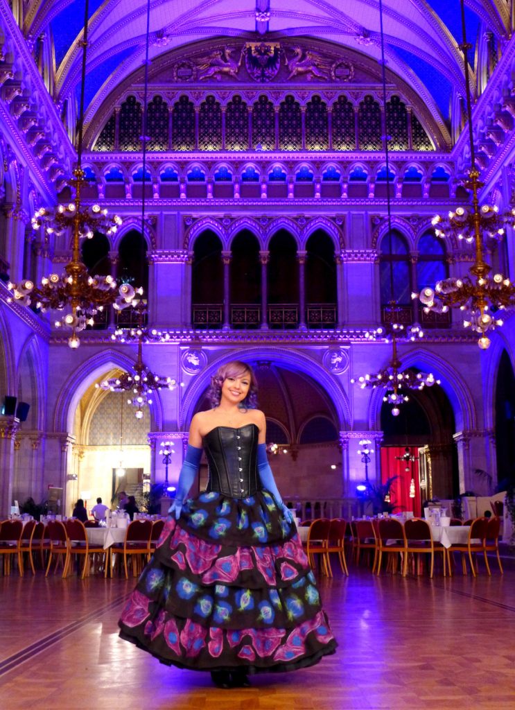
For my microscopy fashion, I spend many hours in a dark microscope room and capture the most beautiful images I can find. I then compile them into aesthetic patterns which are then printed on fabric and used for creating clothes and accessories.

Artwork/Exhibition you are most proud of:

Though it’s hard to pick just one, some events and artworks stand out for their uniqueness. A particular highlight was organizing a “molecular fashion show” to showcase my microscopy dress and other science fashion at the Vienna Ball of Sciences, a popular event celebrating science communication combined with classical traditions.

Beata wearing her microscopy dress, which has a black corset top and multiple tiers on the skirt. The tiers have alternating blue cells and pink cells.
Microscopy Dress (2018) by Beata Mierzwa. A dress representing the beauty of the molecular world! The prints on the skirt are created from real microscopy images of human cells. The blue layers show a collage of dividing cells, with green mitotic spindles segregating the blue DNA into two daughter cells. This ensures that each cell receives the correct set of chromosomes. The purple layers highlight the cytoskeleton made of actin filaments, which provide mechanical support and are essential for cell division and migration.

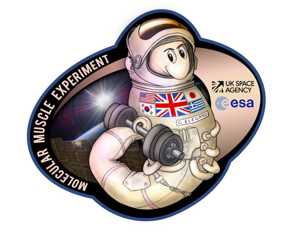
Another one that comes to mind is a mission patch I created for research on spaceflight biology. Researchers sent C. elegans worms into space to study how loss of gravity affects muscle loss and aging. The drawing was highlighted in the media to increase public visibility and used as a way to get students excited about science. After the worms returned from the International Space Station, I was gifted the most unique souvenir—one of their experiment bags that had made the trip to space and back to earth!

C. elegans worm in a space suit and holding a free weight with its tail. The left side of the sticker says "Molecular Muscle Experiment," and the right side has the UK Space Agency and European Space Agency logos.
Space Worm (2017) by Beata Mierzwa. Spaceflight is an extreme environment that causes many negative health changes to the body, among which is space-induced muscle loss. The Molecular Muscle Experiment sends C. elegans worms to the International Space Station to study how zero gravity affects the loss of muscle mass as well as aging.

Which scientists and/or artists inspire and/or have influenced you?

As I was exploring my passions for science art during my doctoral research, I was very fortunate to have the support of both my group leader, Daniel Gerlich, as well as the Institute of Molecular Biotechnology and Vienna BioCenter. Their continuous encouragement throughout my PhD provided an invaluable environment, not only for my science, but also for my artistic adventures.

Two large inspirations for me are Ahna Skop and David Goodsell, both of whom have been pioneers in bringing together art and science throughout their careers. Ahna Skop researches cell division and is also an amazing science artist and communicator. She has been running the Worm Art Show at scientific conferences, where attendees can display their artistic creations inspired by their research. Her story of how she established this art show more than 20 years ago has been a huge inspiration for me. Another wonderful example of a scientist promoting science art is David Goodsell, who creates mesmerizing watercolour paintings based on real structural biology to show the fascinating inner life of cells.

I also admire the artists M.C. Escher and Vladimir Kush, who create surreal worlds through fusing seemingly unrelated elements and imagery.

Panel showing the different stages of cell division with illustrated internal structures.
Complex Organization of Cellular Structures (2017) by Beata Mierzwa. Cellular components are precisely organized down to their very molecules, enabling the cell to perform fascinating processes like cell division. This artwork is a fusion of science and art—real microscopy images of dividing human cells overlaid with drawings that highlight the complex arrangement of DNA, cytoskeletal components, and cellular membranes within the cell division machinery. Illustration for Daniel Gerlich’s lab at Institute of Molecular Biotechnology (IMBA), Vienna.

Is there anything else you want to tell us?

Whatever your dream is, whether it’s science, art, or combining your own unique passions—don’t be afraid to create your own path. Being a scientist doesn’t need to be an all-encompassing definition of who you are; you can have unique hobbies, fantastical ideas, and still be a great scientist as well!

Highlighting the diversity among researchers and painting a more inclusive picture of what it means to be a scientist is our opportunity to challenge current stereotypes and stigmas. I believe that if we make science accessible through creative approaches and encourage multiple passions, then we can inspire a future generation of scientists. This new generation can make enormous contributions with fresh and creative ideas, laying a foundation for innovative research and groundbreaking discoveries.

“Highlighting the diversity among researchers and painting a more inclusive picture of what it means to be a scientist is our opportunity to challenge current stereotypes and stigmas.”

Beata Mierzwa
Different forms of tomato plants on two different paths that converge at the bottom of the image. At the bottom, there is a pair of hands holding modern tomatoes on the vine.
CRISPR Tomatoes (2017) by Beata Mierzwa. Today’s tomatoes are the result of thousands of years of domestication, illustrated by the path on the right towards natural selection of larger fruit size. Independently, a wild tomato strain emerged on the left path, carrying a different mutation that facilitates harvest. A combination of these two beneficial traits would optimize fruit production. However, breeding to cross those “paths” leads to a negative interaction between those natural mutations, resulting in excessive branching and lower tomato yield. This challenge was finally solved using CRISPR/Cas9 to balance the expression of key genes and create an improved tomato plant, as shown on the bottom.
Star map illustration with constellations and drawings of commonly used model organisms and cell types, including a rat, frog, C. elegans, and fish.
Scientific Star Map (2018) by Beata Mierzwa. The intricate patterns of our night sky have inspired many ancient cultures who connected the stars to create constellations portraying myths and legends. This “modern” star map is a scientific take on zodiac signs—showing constellations that form some of the most widely used model organisms and cell types. These model systems have greatly facilitated research and allowed us to discover much of what is known about biology and life today. This artwork was featured on the t-shirts of the ASCB|EMBO Meeting 2018.

For more by Beata Mierzwa, visit her website, Twitter, Instagram, Facebook, or LinkedIn.

Share this Post

About the Author

McKenzie Prillaman

McKenzie is a fledgling science communicator working at the American Association for the Advancement of Science (AAAS). She has a background in neuroscience, and was a research assistant at the University of Virginia and a postbaccalaureate fellow at the National Institutes of Health. After years of thinking she’d become a neuroscience researcher, she discovered her passion for sharing science with others. That finding, in combination with her lifelong dabbling in the arts, led her to write for Art the Science's blog. In her free time, she can be found volunteering with the Smithsonian Associates studio arts classes, trying new foods, and wandering around her home of Washington, D.C. Twitter: @meprillaman