Name: Hannah Warming
Which came first in your life, the science or the art?
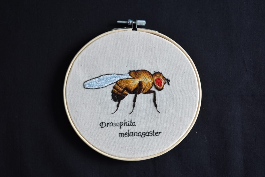
The science came first by many years—my enjoyment of SciArt is a very recent development. I’ve loved biology for as long as I can remember, was obsessed with bugs and animals as a child, and now have a Master’s in Biomedical Science. Since then, I have been working on my PhD in Neuroscience, but discovered a love for embroidery only six months ago.
I always enjoyed painting and drawing but never had much of a natural affinity with it. However, with needlework, I was immediately obsessed and haven’t stopped since.

Which sciences relate to your art practice?
I always said the one field I wouldn’t work in would be neuroscience, until I had a taste of working with brain cells (neurons), and now I can’t imagine doing anything else. Now, I am a neuroscientist and an electrophysiologist in the lab, working with micropipettes to record electrical signals from cells.
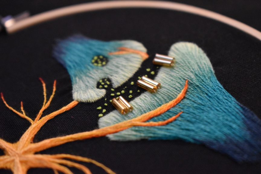
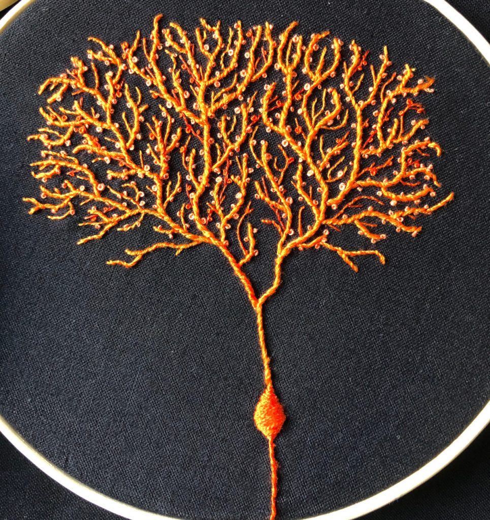
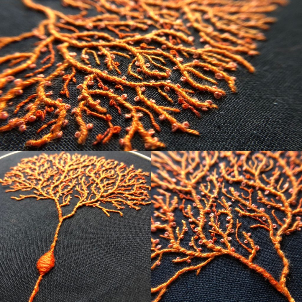
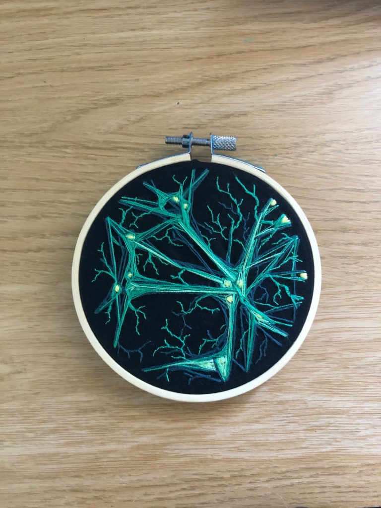
When I am recording data with electrophysiology, I don’t just look at the cells, but I touch them and manipulate them. I will press the pipette against the cell membrane and gently break through to record currents from within the cell. In this work, you can almost feel the elasticity of the cell membranes when you pull a pipette away. These physical properties of the cell membrane fascinate me. I grow primary neuron cultures and can’t get enough of the fibrous texture and intricacy of the axons and dendrites that these cells grow.
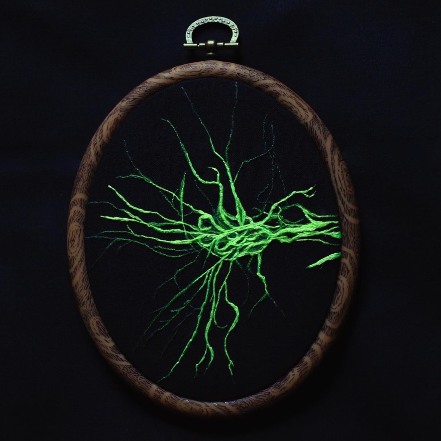
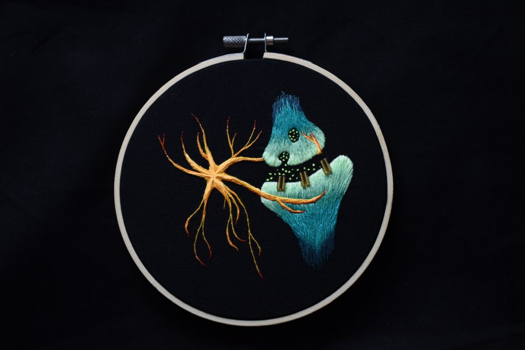
Working so intimately with neurons and “feeling” that texture of the cells makes it very natural for me to recreate them with thread—it is the perfect medium to represent them in a tangible form outside of the brain. I will never get tired of stitching neurons, and I hope I can work with them for many years to come.
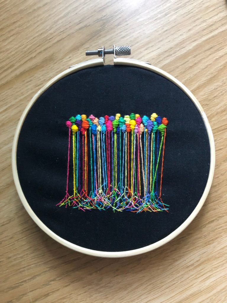
Seeing the patterns that occur naturally in the brain, like the cell layer organisations in the hippocampus or cerebellum, is a huge inspiration for my art. I want to share with people the beauty of the brain, from the perspective of someone who works with it on a single-cell level. In the process, I am hoping to inspire a few people to enjoy neuroscience or even to pursue a career in research.
“Working so intimately with neurons and ‘feeling’ that texture of the cells makes it very natural for me to recreate them with thread—it is the perfect medium to represent them in a tangible form outside of the brain.”
Hannah Warming
What materials do you use to create your artworks?
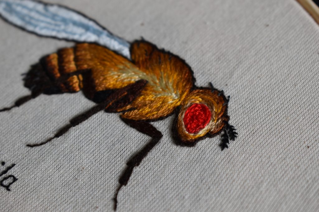
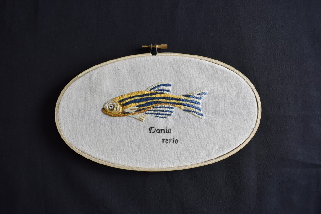
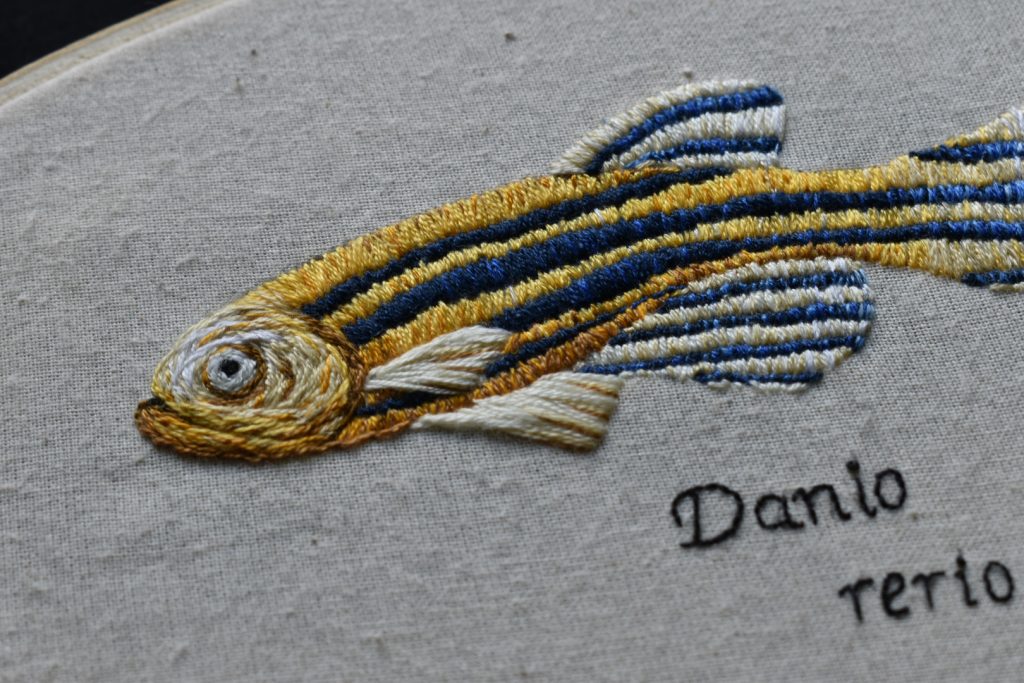
I mainly use standard cotton threads on calico fabric, but have recently started trying out light reflective threads and beads to catch the light in new ways. I plan to learn new techniques to incorporate 3D designs in future, as well as embroidery on clothing (well, lab coats to be specific!).
Beyond learning a few basic stitches and knots at the beginning, I tend to go by instinct—I have the idea in my head of the texture I want to create, and work the threads until I achieve it. I recommend embroidery to anybody who is interested—you need very little to get started but sewing needles, thread, and a bucketload of patience!
Artwork/Exhibition you are most proud of:
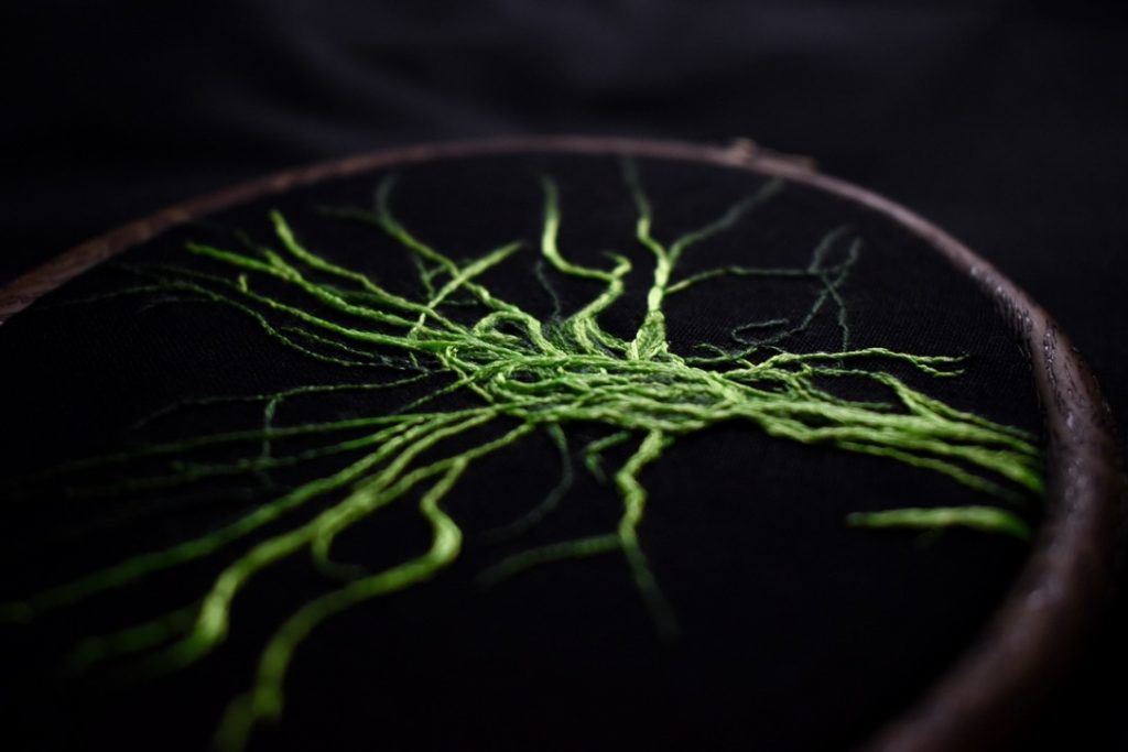
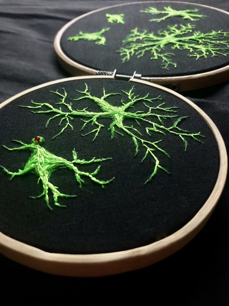
My Purkinje cell embroidery in August 2020 had an amazing reception on Twitter—I wasn’t expecting it at all! It has now been shipped from the UK to the USA, but is also on display digitally on the ConciliARTe online exhibition. This piece took away a lot of my “imposter syndrome” and showed me that perhaps I do belong in the science art community after all. The online exposure also allowed me to discover other likeminded scientists and interact with the amazing online community of artists, which has been really supportive.
“This piece took away a lot of my ‘imposter syndrome’ and showed me that perhaps I do belong in the science art community after all.”
Hannah Warming
Which scientists and/or artists inspire and/or have influenced you?
One of the first people I saw doing neuroscience embroidery was Lauren Hewitt—her detailed neurons and hippocampal diagrams are incredible. I found more creators, such as Laura Bundesen, firstly on Instagram and then on Twitter. They were making fibre art inclusive and slowly grew a collection of inspiring artists who can draw, paint, and create art that really shows a passion for their field of interest. I highly recommend checking out @Krakenkreationscath on Instagram—her anatomical embroideries are breath-taking.
A final mention goes to two artists I met through work, Barbara Touati-Evans and Susan Merrick, whose latest project is “Detangling the Knots.” It uses fibre arts and performance to represent the “tangles” in the brain that cause Alzheimer’s disease and reflect on its impact. They visited us in the lab to see our research hands-on, and really helped me to see the beauty I had been taking for granted in the work I do.
Is there anything else you want to tell us?
My embroidery is for charity! Donations are at an all-time low for many charities due to the ongoing pandemic and the financial trouble it has caused across the globe. I work closely with teams researching dementia, and am part of the Alzheimer’s Research UK network. So when I took up embroidery, I decided 50% of any sales I made would go straight to ARUK—every purchase donates money to help find a cure for dementia.
I encourage anyone with a curiosity for art to embrace it, and see where it takes you—worst case scenario, you have fun!
For more by Hannah Warming, visit her Instagram or Twitter.
Share this Post